Blastocysts and hCG----Objective 78
I would just like for you to know that I am laughing at myself right now. Firstly because I just realized I am on my last objective and secondly because I just read the last objective and for the life of me could not "recognize that blastocyst secrete human gonadotropin". I am so exhausted and mentally fried that I could not figure out what in the world is human gonadotropin! And then it its me...duuuuhhhh...its the same hormone that you test for when you find out your pregnant!! Which is the same hormone that prolongs the life of the fetus...At this point I am glad I am laughing at myself and not crying.
The Birds and the Bees---Objectives 71, 72
Discovery Health has a pretty funny video about conception. Before watching this video I did not know that there is an enzymatic cap, on the head part of the sperm, that allows the sperm to penetrate the egg.
Zygote, Morula Blastocyst...Oh My!---Objective 76

Morula is the process where the zygote begins to divide and multiply into many cells.
A blastocyst begins at day 5 in mammals after fertilization.
This all occurs during the first week of being conceived
Individual Responsibility---Objective 77
In A & P 210 with Dr. Middleton, we had to do a community service project that involved the class donating blood to a local blood bank. At first, I was a little nervous because I have never given blood before and I had heard that the needles were HUGE! Well push came to shove (my procrastinating habits came into play) and I finally decided to get it over with (at the very last minute). To tell you the truth it wasn't that bad. The needle WAS huge and the nurse said I have pretty deep veins, so there was some pain involved, but hey I've given birth to two VERY big headed daughters so I was able to deal with the pain. Anyways, to make a long story short, I have given blood ,every 2 months or so, since then. If it was not for Dr. Middleton and that dreaded project I would have never realized the importance of giving blood.
Spermatogenesis and Oogenesis---Objective 73, 74
This video compares spermatogenesis and oogenesis. To me it is helpful to see the mitotic division of each one side by side so you can get a better comparison of the two.
The Liver----Objective 59
This website made learning about the microscopic anatomy of the liver easy. I was able to enlarge the microscopic picture of the liver and color/label each structure. It was also helpful because it gave a description of each structure.
Erythropoietin and Red Blood Cell Production---Objective 54

This diagram helped me understand the regulation of red blood cells. The flow of things is pretty easy to understand now. Oxygen levels decrease, kidneys produce erythropoietin which stimulates production of RBCs in bone marrow, RBCs carry oxygen to body. And the loop continues...
Spirometer Use---Objective 51
Although the physioex exercise were long and miserable, they really did help furthering my understanding how the different systems work. Here is a snipet from the respiratory physio-ex showing the use of a spirometer.


Oxidative Phosphorlation
I do remember this from Bio-100. Here is a diagram that helped to refresh my memory.
 Oxygen is crucial for oxidative phosphorylation because it is the last electon carrier.

Carbon Dioxide and Ventilation---Objective 48
Here is a snipet from our Respiratory Reciation:


Participating in the recitations did help pull all of the systems together as a whole. I also think that researching the the different systems to answer these questions helped us meet some of the objectives needed for this class.
Cardiovascular disease---Objective-30
Americanheart.org shows some unbelievable statistics about deaths caused by cardiovascular disease. My guess that the statistics are so high is due to that cardiovascular disease is classified as a large array of diseases including high blood pressure, strokes...etc
Genetic Disease---Objective 26
4 types of genetic diseases:
Monogenic (single gene) ex: sickle cell anemia, cystic fibrosis, Aicardi Syndrome, Huntington’s disease
Polygonic (multiple genes) ex: Alzheimer, diabetes, obesity and arthritis
Chromosomal ex: Down Syndrome
Mitochondrial ex: cause deformities
Monogenic (single gene) ex: sickle cell anemia, cystic fibrosis, Aicardi Syndrome, Huntington’s disease
Polygonic (multiple genes) ex: Alzheimer, diabetes, obesity and arthritis
Chromosomal ex: Down Syndrome
Mitochondrial ex: cause deformities
Blood Clotting Process---Objective 22
A video that shows the blood clotting process. It really helped watching how the RBCs, platelets, and clotting agents work together to form a clot.
Technology---Objective 19
This website (scroll to the bottom) gives a timeline of historical events in anatomy and physiology. I did not know that the first human dissection was done in Greece in 500 BCE.
Lactation---Objective 18
A great chart showing the flow of hormones and the stimulus that initiate lactation. It is really easy to follow this diagram to understand the effects of hormones and stimuli.


The Menstural Cycle---Objective 16, 75
This website was very informational on the effects of estradiol and progesterone on menstruation. It included easy to understand text and charts. Whoever knew Aunt Flow was so complex...haha!
Endocrine Sytem and Homeostasis---Objective 11, 12, 13, 14


This table made it easy to identify the classes of hormones
Venous Valves and Varicose Veins---Objective 31

In this picture you can clearly see that normal vein valve operation only allows blood to flow one way (towards the heart). But in varicose veins the valve is damaged allowing blood to flow both toward and away from the heart, causing the veins to become enlarged.
Inspiration and Expiration---Objective 46, 49, 50
I like videos that dumb things down...inspiration and expiration for dummies. I thought it was helpful to see the changes of barometric and aveolar pressure.
Neuronal Control Of Inspiration and Expiration:
 This picture was actually taken from our Anatomy 211 textbook. Although it was difficult to understand at first, the further I read about the medullary and pontine respiratory centers, the easier it was to understand the diagram and also how the neurons fire inspiration and expiration.
Neuronal Control Of Inspiration and Expiration:

Acidosis or Alkalosis?---Objective 53, 56
Have to give credit to the Super Nurse in my life, my mom, for this one. She explained respiratory and metabolic alkalosis and acidosis in a way that made it really simple.
Remember ROME---> Respiratory-Opposite Metabolic-Equal
If your pH is low and your PCO2 (*partial pressure of carbon dioxide) is high: high/low is opposite. So its respiratory, and you know its acidosis because the pH is low.
If your pH is high and your HCO3 is high. high/high is equal, so its metabolic. and you know its alkalosis because the pH is high
*partial pressure is the pressure exerted by one gas in a mixture
Respiratory Anatomy---Objective 45
Again my kids come to the rescue. Coloring...who would of thought that would help me study!
My picture of the respiratory system that I would color:

My picture of the respiratory system that I would color:

Edema--Objective 55
I have always thought of edema simply as swelling. But the more that I have learned about the different systems of the body I've realized that causes and/or effects of edema occur throughout the organs. The most common being heart, liver and lungs. Here I was thinking edema only happened if you were pregnant or ate too much salt...boy was I wrong!
For example, a patient with heart disease. Poor cardiac function can cause a decreased volume of blood pumped out by the heart. The decreased cardiac output is responsible for a decreased flow of blood to the kidneys. The kidneys can sense that there is a reduction of the blood volume in the body. To counter act the loss of fluid, the kidneys retain salt and water. The kidneys are then fooled into thinking that the body needs to retain more fluid volume but, in actuality the body is already holding too much fluid. This fluid ultimately results in the buildup of fluid within the lungs, which can cause shortness of breath. This accumulation of fluid in the lung is called pulmonary edema. At the same time, there is also an accumulation of fluid in the legs causing edema.
WOW! It's amazing how all the systems coincide with each other!
Kidney Function---Objective 52, 62, 66
Today in lab when Mrs. Gess was discussing the function of the kidneys, I was so lost. (Sorry Mrs. Gess but the drawing on the board was just not doing it for me!) There is a website that I've looked at before for 210 that I have found very useful and I was grateful that they had a video on the kidneys. This video dumbed things down for me and it was helpful to see how things travel through the kidney.
Kidney structure---Objective 63

Vasoconstriction and Vasodilation---Objective 36
This one is pretty easy to remember. Its all in the words!
Vasoconstriction = veins constrict
Vasodilation = veins dilate

Vasoconstriction = veins constrict
Vasodilation = veins dilate

Protection Against Pathogens---Objective 38, 39, 40, 42, 43, 44
A good story to remember how the lymphatic system protects the body from pathogens.
The white blood cells act as the body's military, the body is a huge battle ground. The war goes on forever. We are of course rooting for the WBC Army, but the Foreign Microbe Armies are impressive in their own right.
The WBC army bases are (where WBC's are produced) in the land of the Red Bone Marrow. These are well protected locations in large bones for the WBC to gather and plan their attack (sternum, long bones of the arms & legs, hip bones, scull, vertebrae). When they're ready and pass basic training they all take the Blood and Lymphatic Rivers to get anywhere in the world of the Body.
The WBC army has spies and scouts: chemicals that are released into Tissue Land from the invader microbes (pathogens). These chemicals signal the WBC's, telling them location, type of microbe, and the invaders numbers. Famous scouts are our brothers Histamine, Complement, Kinins and Leukotrienes. I always like to imagine microbes having a big loud obnoxious party after they pass the Great Skin Barrier (Body's first defense) and set up camp inside. They're jerks like that. They trash the neighborhood (damage nearby tissue).
Neutrophils are the first responders - the marines. They're tough and scrappy (they have granules in the cytoplasm and a multi-lobed nucleus). These guys are large in number - around 70% of the entire WBC army. They fight bacteria and fungal microbes the best. They show up early while the microbe invasion party is still going strong and begin to bust everything up. Acute inflammation is evidence of their fight. Pus litters the battlefield (a combo of dead neutrophils, microbes, and fluid and tissue from the area).
Next the Monocytes come and and clean up the area after the fight: "The Cleaners." They're different from the neutrophils - only 1 nucleus and no granules. They collect evidence of the invaders and send it to the T-cell division (lymphocytes who will remember the information for future attacks). After they're done in damaged Tissue Land they go back to the Lymphatic River. They can transform into macrophages.
Macrophages are big tanks that can kill larger microbes that have made it past the neutrophil battle. Like the monocyte, macrophages have a single nucleus. Marcrophages set up camps throughout the body waiting for any invader microbes that may have made it past enemy lines. They have different names for different camps throughout the body: ie Dust Cells in Lungs-ville, Kupffer cells in Liver-town. They like to set up road blocks in sinus cavities and lymph nodes where they can stop any microbes trying to get further past enemy lines.
Basophils, scrappy like the neutrophils (bi or tri lobed nucleus, full of granules) and show up and increase the inflamation. The bomb makers. They especially like to bomb allergies.
Eosinophils are also pretty scrappy like the neutrophils (bi-lobed nucleus and full of granules) and they arrive early to inhibit all the inflammation. The bomb squad maybe. They increase in numbers when the microbes are parasites or allergies because that's the fight they like to get in on.
A lymphocyte called the Natural Killer Cell helps out by recognizing human cells that have been invaded by viruses or tumor cells trying to establish bases past enemy lines. NK's are bad asses that notice the markers on the cells, then it kills the ones that indicate virus or tumor - no questions asked.
All these cells are part of the Innate Immune System - they're non-specific - they'll fight any pathogen that tries to set up shop. The Adaptive Immunity System is set up for repeat offenders. Remember the T-cell (friend of the monocyte) above? Along with the B-cell lymphocyte they remember pervious invaders and activate attacks accordingly. It's a big old war going on in there. All day, every day.
Using this analogy helped me meet most of the objectives of the lymph system. What a fun way to learn and teach this system!
Structures and Functions of Lymphatic System---Objective 37
I really hate just staring at a book for hours trying to memorize different names and placements for different structures. Its a good thing that I live in the age of the Internet. I found a website that has matching tutorials for: components of the lymphatic system , structure of a lymph node, and routes of drainage of lymph.
Oh boy do these help!
A mnemonic I made up to remember the function of the lymphatic system:
The Flu Is Even Dead---> Transports nutrients, Fluid and protein balance, Immunity, Excretion of waste, Digestion( aides in absorption of fat)
Oh boy do these help!
A mnemonic I made up to remember the function of the lymphatic system:
The Flu Is Even Dead---> Transports nutrients, Fluid and protein balance, Immunity, Excretion of waste, Digestion( aides in absorption of fat)
Blood Pressure---Objective 35, 65

My interpretation of the above diagram...
blood pressure falls---> the kidneys release renin into the bloodstream.
Renin ( secreted by juxtaglomerular cells) splits angiotensinogen
Angiotensin I--->is split into pieces by angiotensin-converting enzyme (ACE).
Angiotensin II causes the arterioles to constrict, increasing blood pressure.
Angiotensin II triggers the release of aldosterone from the adrenal glands and antidiuretic hormone from the pituitary gland.
Aldosterone causes the kidneys to retain sodium and excrete potassium.
The sodium causes water to be retained, thus increasing blood volume and blood pressure.

Naming the Arteries and Veins---Objective 23, 33, 34
Its funny how I've realized that the best way for me to learn and study is the same way I study with my kids. I play games! Here is a great game that has come in handy when I've need to study for lab practicals.
A good picture of major veins:
 Superior vena cavae (or precavae) drain the anterior regions of the body
Inferior vena cava (or postcava) drains the posterior portion of the body
A good picture of major veins:

Inferior vena cava (or postcava) drains the posterior portion of the body
Gonadotropins---Objective 15
Gonadotropins = stimulate gonads
 This diagram is pretty easy to interpet. The hypothalamus synthisizes and releases gonadotropin-releasing-hormone. This hormone stimulates secretion of LH and FSH, which in turn stimulates gonadal secretion of the sex steroids testosterone, estrogen and progesterone. I really like this diagram, I wish our lab manual had included this diagram.
Blood Types---Objective 21
The Blood Typing Game: to play this game you have to match the donor to the recipient. It helps to learn the correct matches because an incorrect match can be fatal.
In lab we had to experiment with blood typing to determine each type of blood. This website really contributed to the blood typing we did in lab. It was interesting to see the blood coagulate when the anti-A and anti-B antibodies were added.
In lab we had to experiment with blood typing to determine each type of blood. This website really contributed to the blood typing we did in lab. It was interesting to see the blood coagulate when the anti-A and anti-B antibodies were added.
Knowing the Muscles
I wish I had a picture of my husband working out to his P90x videos, but my way of helping me to learn the names of the different muscles is to tell him which muscles he is using while he works out. Not only am I learning but I am also making him think that I am really smart...haha!
Applying Thinking Skills and Solving for Unknowns--Objective 2, 3, 4, 7,9, 61
The Food Nutrient Analysis really put our critical thinking skills to work. In this lab, we went from station to station testing the presence of sugar, starch, lipids, vitamin C, and proteins in various solutions. At the last station, we had to test two unknown solutions for the presence of organic compounds. This came a little easy for me; this is what I used to do at my job as a chemical lab technician.
 Here is my lab partner, Nancy, testing for the presence of sugar.
The Anatomy of the Heart---Objective 24, 25
I was so lost in lab when we were learning about the structures of the heart! In complete desperation I happen to find this fun and interactive game that helped me learn the anatomy of the heart!


Compare and Contrast Bond Types---Objective 10
Learning about the different types of chemical bonds was tough. Luckily, I used to work in a chemical laboratory and have previously been exposed to different types of bonding, which helped whe we were learning more about them.
I did find a tutorial that has an itroduction, animation and even a quiz that reinforced everything I had learned in previous classes ( Bio-100) and labs.
I did find a tutorial that has an itroduction, animation and even a quiz that reinforced everything I had learned in previous classes ( Bio-100) and labs.
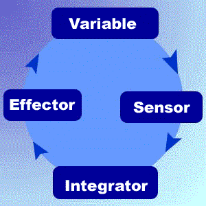
Understanding Homeostasis---Objective 1
Whenever I think about homeostasis, the first thing I refer to is the example of the thermostat.
Negative Feedback = thermostat's response causes temperature decrease to reverse and become a temperature increase (
Positive Feedback = the thermostat's response to a dropping temperature was to switch off the furnace or to switch on the air conditioner

Negative Feedback = thermostat's response causes temperature decrease to reverse and become a temperature increase (
- shivering in response to cooling of body during cold weather
Positive Feedback = the thermostat's response to a dropping temperature was to switch off the furnace or to switch on the air conditioner
- increased labor contractions stimulated by oxytocin (OT) hormone
 |
| Room Control |
 |
| Feedback Loop |
 |
| Human Body Regulation |
The fish bowl example shows how each organ system maintains homeostasis.

Also a cool video about how the body maintains water and glucose balance.
Heart Valves---Objective 29, 32
I had a hard time remembering the order of blood flow through the heart valves until I watched this video from the Heart Lung and Blood Institute. It helped to hear someone explaining and pointing to each part of the heart as the blood entered the heart.
All Physicians Take Money=Aorta, Pulmonary, Tricuspid, Mitral...Thanks Nancy for that one!
Blood flow during systole and diastole...
All Physicians Take Money=Aorta, Pulmonary, Tricuspid, Mitral...Thanks Nancy for that one!
Blood flow during systole and diastole...
A co-worker that my mom works with, Dr. Assorgi, with told me about a website, http://www.medicalvideos.us/, that had a ton of videos about every organ system we were learning about. Thanks to him I found the video above that helped me understand systole and diastole.
Heart Sounds and Conducting System Objective 27, 28, 29
This animation, although not very anatomically correct, gives a nice visual of how the S-A and A-V work. What makes it even better and helpful is the step-by-step process the animation shows.
Another animation of the heart during systole and diastole. I thought this animation was just awesome. You can see everything working through all the phases of systole and diastole.
Although kind of long, the video below lets you listen to various heart sounds, which made it helpful to compare and contrast the different sounds.
Adrenal Gland and Stress---Objective 17
 (a picture of me while trying to complete this portfolio)
I found this video online that helped me understand that stress can cause an excess release of cortisol which can lead to being overweight. The video was able to break it down to layman's terms to relate to what we are learning in class.
Following the River of Life---Objective 20, 64
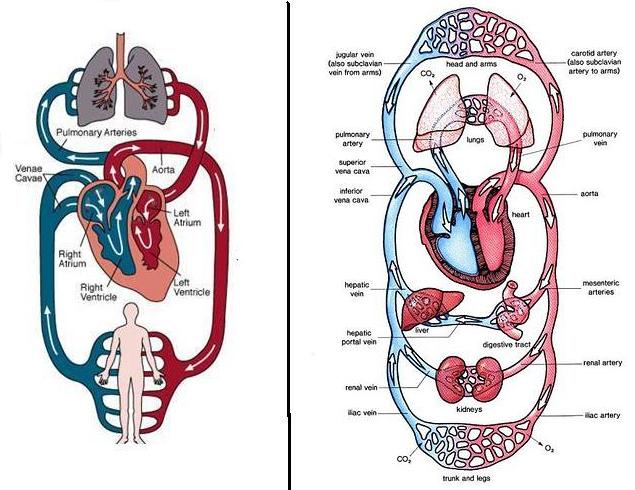
After looking at these for what seemed like forever and still not getting the flow correct, I started printing them out in black and white and coloring the pictures. Coloring the veins blue for deoxygenated blood and the arteries red for oxygenated blood helped me learn the pathway of the blood. Arteries=Away
Also getting this song(<-- click there to listen) stuck in your head during a lab practical doesn't hurt either! The lyrics:
Pup, pump, pumps your blood
The right atrium's where the process begins,
Where the CO2 blood enters the heart.
Through the tricuspid valve to the right ventricle
The pulmonary artery and lungs.
Once inside the lungs it dumps it carbon dioxide
And picks up its oxygen supply
The it's back to the heart through the pulmonary vein
Through the atrium and left ventricle.
Pump, pump, pumps your blood.
The aortic valves where the blood leaves the heart
Then it's channeled to the rest of the bod
The arteries, arterioles, and capillaries too
Bring the oxygenated blood to the cells
the tissues and the cells trade of waste and CO2
Which is carried through the venules and the veins
Through the larger vena cava to the atrium and lungs
And we're back to where we started in the heart."
During the lab practical last week I actually caught myself tapping my foot at the beat of this song...but hopefully it helped me enough to get me through that test!
Endocrine or Exocrine...Can you tell the difference?---Objective 5, 6
 |
| Ted Striker putting his exocrine glands to work! |

*Endocrine=In*

Subscribe to:
Comments (Atom)